piVector Designer

Optimized for high-quality AAV production and vector design
Overview
Plasmid design
Welcome to piVector Designer, this free online DNA construct design tool will simplify your plasmid design work. Developed with researchers in mind, this cutting-edge plasmid design tool streamlines the conceptualization of DNA constructs, making complex tasks more accessible and efficient.
Equipped with a vast library of plasmid backbones that are suited for a wide range of applications, the piVector Designer is your go-to resource for viral vector design. Our base library includes numerous AAV and lentivirus vectors that have been rigorously verified for effective viral packaging to enable worry-free vector design. Additionally, we are excited to announce the upcoming availability of vectors for protein expression, gene editing, shRNA, and cloning to further broaden our support for your plasmid construction needs.
Research Optimization Program
Academic Discovery Engine
Accelerate your research from design to delivery with academic exclusive offers
gift icon

Free gene synthesis and cloning + Win gift card for your feedback!

  • Free gene synthesis for human and mouse ORFs or gene elements from our database.
  • Free cloning with any plasmid prep or AAV/lentivirus packaging order.
  • We value your feedback! Click the “Feedback” gift box icon on the right side of the screen while using or after using piVector Designer. After completing a short survey, you’ll be directed to a lottery wheel for a chance to win a prize!

  • *Offer cannot be combined with other discounts
    PackGene reserve the rights to interpret the promotion

How To Use

Engage in an intuitive design experience by selecting a backbone and incorporating your gene sequence or choosing from pre-loaded elements. Our plasmid design tool makes constructing a functional vector as straightforward and enjoyable as assembling building blocks. You can tailor your design to include various expression cassettes for regulated gene expression, fluorescent labeling, enhanced gene expression through regulatory elements and poly A signals, and the delivery of gene editing machinery into target cells.

Our built-in error-detection script meticulously verifies your design to ensure accuracy in reading frames and translation stop positions. With the PackGene Vector Design error-detection script you can have confidence in the quality of your plasmid construction.

Progressing from design to realization is seamless with PackGene Vector Design. Our integrated ordering process includes gene synthesis, cloning, AAV or lentivirus packaging, among other services. Our aim is to streamline the transition from vector construction to application. You may also request additional plasmids or analytical tests to support your broader research, development, or preclinical study objectives with ease.

Key Benefits of piVector

Reliable Backbones

Enhances production efficiency and boosts AAV Yield.

multitypes icon

Proven Performance

RC plasmids tested for consistency

icon19

Extensive Gene Elements

Promoters, regulators, reporters and ORF from NCBI GenBank

icon23

User-Friendly Design

Intuitive interface for effortless vector design

icon20

Digital Management

Secure, easy vector tracking and reordering

icon4

Cost-Effective

Pre-synthesized elements save time and fees

Performance

piVector as a GOI Plasmid Backbone Greatly Enhances AAV Production Yield

AAV data

piVector as a GOI Plasmid Backbone Significantly Reduces Residual Plasmid DNA in AAV Production

Residual plasmid DNA ssAAV
*Residual plasmid DNA in AAV is measured using qPCR, which quantifies DNA by targeting the AAV ITR and plasmid ori regions, ensuring sensitive and accurate assessment of purity and quality.
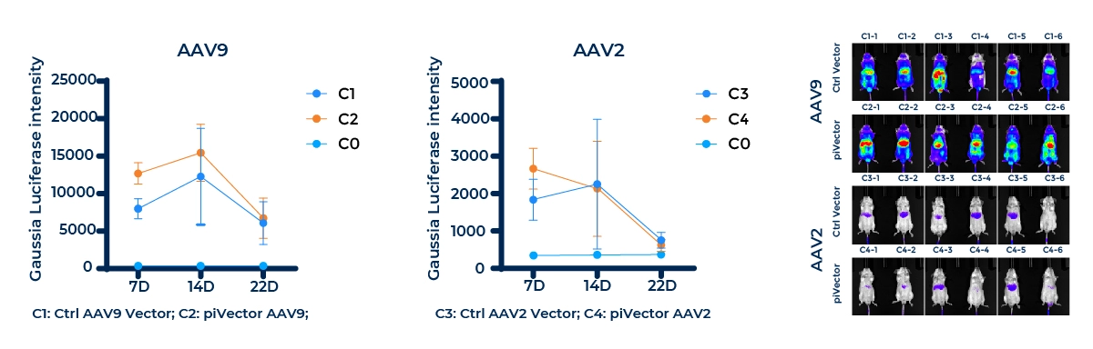
How piVector Designer Revolutionizes Plasmid Design
Comprehensive Design
Extensive Library
Professional Interface
Streamlined Ordering
Information & Technical Support

PackGene is dedicated to propelling your research forward. For more information on how piVector Designer can enhance your plasmid design and gene editing projects, contact us at info@packgene.com

Resources

icon21

piVector Designer Flyer

icon21

Investigating Opioid Peptides: AAV Vector-Delivered Fluorescence Sensors for Studying Opioid Dynamics

icon21

How to Optimize the Design of AAV and Lentivirus Transfer Plasmids

FAQ

What are the components of plasmid vectors?

Plasmid vectors are indispensable in genetics and molecular biology, serving as vehicles to insert, manipulate, and transfer genes within organisms. They typically contain key components like the origin of replication (ori), promoter regions, open reading frames (ORFs), regulatory elements, a polyadenylation (poly A) signal, multiple cloning sites (MCS), and antibiotic resistance genes. Choosing the right combination of these elements is crucial for the success of your research, as each one plays a specific role in plasmid functionality.

What bacteria strains do you use for cloning and plasmid preparation?

We use the strains below depend on different applications:

1. DH5α

  • Genotype: F–, φ80dlacZΔM15, Δ(lacZYA-argF)U169, recA1, endA1, hsdR17(rK–, mK+), phoA, supE44, λ–, thi-1, gyrA96, relA1
  • Applications: Commonly used for general cloning purposes and blue/white screening. Its mutations in recA and endA enhance plasmid stability and transformation efficiency, making it a go-to strain for many cloning applications.


2. Top10

  • Genotype: F–, mcrA, Δ(mrr-hsdRMS-mcrBC), φ80lacZΔM15, ΔlacX74, recA1, araD139, Δ(ara-leu)7697, galU, galK, λ–, rpsL(StrR), endA1, nupG
  • Applications: Suitable for general cloning and blue/white screening with high transformation efficiency. It is often preferred when maximizing the number of transformants is critical.


3. Stbl3

  • Genotype: F–, mcrB, mrr, hsdS20(rB–, mB–), recA13, supE44, ara-14, galK2, lacY1, proA2, rpsL20(StrR), xyl-5, λ–, leu, mtl-1
  • Applications: Ideal for cloning vectors with repetitive elements, such as long terminal repeats (LTRs) in lentiviral plasmids. The recA mutation minimizes recombination, enhancing the stability of complex constructs.


4. XL-10

  • Genotype: TetR Δ(mcrA)183 Δ(mcrCB-hsdSMR-mrr)173 endA1 supE44 thi-1 recA1 gyrA96 relA1 lac Hte [F´ proAB lacIqZΔM15 Tn10 (TetR) Amy CamR]
  • Applications: Optimized for high-efficiency transformation, particularly for large or methylated DNA constructs. It is also suitable for blue/white screening and for cloning unstable or toxic sequences due to the recA mutation.


5. NEB Stable

  • Genotype: F´ proA+B+ lacIq Δ(lacZ)M15 Tn10 (TetR) endA1 recA1 hsdR17(rK– mK+) glnV44 λ– thi-1 gyrA96 relA1 spoT1
  • Applications: Designed to maintain unstable plasmids that might recombine or degrade in other strains. It is ideal for cloning large or recombination-prone plasmids, offering good yield and stability for long-term propagation.

How is a plasmid constructed based on your design?

We use seamless cloning to construct most plasmids.

Seamless cloning is a method that allows precise insertion of DNA fragments into plasmid vectors without adding extra nucleotides at the junctions, a common issue with traditional restriction enzyme methods. This is crucial for protein expression, as even small additional amino acids can affect protein function. There are various approaches to seamless cloning, including techniques like overlap extension PCR or commercial kits.

Key Steps in Seamless Cloning:

  • Creation of Overlapping Ends: The DNA fragments are generated through PCR with ends that overlap with each other or the plasmid. These overlaps are designed into the primers used for amplification.
  • Annealing of Overlapping Ends: The complementary overlapping regions hybridize when mixed together.

Extension and Ligation:

  • A polymerase may extend the annealed fragments, filling in gaps.
  • DNA ligase then seals the nicks, or a commercial system may combine both enzymatic steps in a single process.

Requirements:

  • DNA Fragments with Overlaps: These fragments, usually created by PCR, must have complementary sequences for hybridization.
  • Polymerase: A polymerase is needed for filling in gaps, if required.
  • DNA Ligase: Ligase seals the nicks unless using a system that combines all steps.

Pros and Cons:

  • Pros:
    Enables precise, in-frame gene insertions without unwanted nucleotides.
    No need for restriction enzyme sites, giving more design flexibility.
    Simplified primer design.
  • Cons:
    Primer design requires precision for correct overlap and orientation.
    Efficiency can depend on factors like fragment size and sequence complexity.
    Commercial kits can be expensive.

Tips and Tricks:

  • Optimal Overlap Length: Overlaps of 15–25 nucleotides typically provide efficient annealing, though larger fragments may require longer overlaps.
  • High Purity DNA: DNA fragments should be pure, often achieved by gel purification post-PCR, for optimal results.
  • Control Reactions: Always include controls (like no-insert controls) to detect potential background noise or unwanted ligation.

What plasmid quality control (QC) tests are performed?

For research-grade plasmids, we perform the QC tests listed below.

  • Appearance: A visual inspection of the plasmid solution to assess its clarity and the absence of particles or discoloration.
  • A260/280: Measures the purity of plasmid DNA by comparing absorbance at 260 nm (nucleic acids) and 280 nm (proteins). A ratio of ~1.8 indicates pure DNA.
  • Homogeneity by Agarose Gel: Assesses the uniformity of plasmid DNA by running it on an agarose gel to ensure consistent molecular weight and the ratio of supercoiled plasmids.
  • Restriction Analysis: Verifies the identity and integrity of plasmid DNA by cutting it with specific restriction enzymes and analyzing the resulting fragments via gel electrophoresis.
  • Endotoxin by LAL: Detects bacterial endotoxins in plasmid preparations using the Limulus Amebocyte Lysate (LAL) assay, ensuring plasmids are safe for sensitive applications.


Additionally, upon request, we offer extra QC tests, which are also included in our preclinical plasmid quality control.

  • Homogeneity by HPLC: Uses High-Performance Liquid Chromatography (HPLC) to evaluate the uniformity and purity of plasmid DNA, and measure the ratio of supercoiled plasmid DNA.
  • Residual RNA by SYBRGold: Quantifies any remaining RNA in the plasmid preparation by staining with SYBRGold and analyzing fluorescence intensity.
  • Residual E. coli DNA by qPCR: Detects and quantifies residual E. coli genomic DNA in plasmid preparations using quantitative PCR (qPCR).
  • Bioburden Testing by Direct Inoculation: Assesses the microbial contamination level of the plasmid preparation by inoculating samples in growth media and monitoring for microbial growth.
  • Sequencing by Sanger: Confirms the accuracy of the plasmid sequence by using Sanger sequencing to verify the inserted DNA or the entire plasmid.
  • Residual Host Protein by ELISA: Detects any leftover host proteins in plasmid preparations using an ELISA assay specific to E. coli proteins.
  • Mycoplasma Contamination by qPCR: Screens for mycoplasma contamination in the plasmid sample using sensitive qPCR techniques.
  • pH by Potentiometry: Measures the pH of the plasmid solution to ensure it is within the acceptable range for stability and application.
  • Residual Kan by ELISA: Detects any remaining kan antibiotic from the plasmid selection process using an ELISA assay.
  • Sterility: Confirms the absence of viable microorganisms in the plasmid preparation, ensuring it is sterile and suitable for sensitive applications.
  • Osmolality: Measures the osmolality (concentration of solutes) in the plasmid solution to ensure it is within acceptable limits for biological compatibility.
Q
download_piVector-Designer-flyer